anatomy of the eye
Anatomy of the Eye
Anterior Chamber – the area located between the iris and the cornea.
Choroid – the thin, blood-rich membrane that lies between the retina and the sclera and is responsible for supplying blood to the retina.
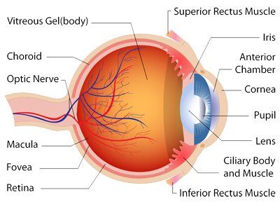
Ciliary body – the part of the eye that produces aqueous humor. Near the ciliary body is the trabecular meshwork, which is where an iStent® for glaucoma is implanted.
Cornea – the clear, dome-shaped surface that covers the front of the eye. Refractive surgeries such as LASIK and PRK are performed on the cornea.
Anatomy of the Eye
Inferior rectus muscle – one of six extraocular muscles that control eye movement.
Iris – the colored part of the eye. The iris is partly responsible for regulating the amount of light permitted to enter the eye.
Lens (also called crystalline lens) – the transparent structure inside the eye that focuses light rays onto the retina. The posterior capsule surrounds the lens. This is where a cataract develops.
Macula – the focusing portion of the eye that allows us to see fine details clearly. Macular degeneration affects this area.
Optic nerve – a bundle of nerve fibers connecting the retina with the brain. The optic nerve carries signals of light, dark, and colors to the area of the brain (the visual cortex), which assembles the signals into images (i.e., our vision). Glaucoma damages the optic nerve.
Punctum – these tiny openings in the margins of the eyelids help collect tears produced by the lacrimal glands. These openings are where a punctal plug would be inserted to help with dry eye.
Pupil – the opening in the middle of the iris through which light passes to the back of the eye.
Retina – the light-sensitive nerve layer that lines the back of the eye. The retina senses light and creates impulses that are sent through the optic nerve to the brain.
Superior rectus muscle – one of six extraocular muscles that control eye movement.
Vitreous body – a clear, jelly-like substance that fills the back part of the eye. “Floaters” occur in the vitreous.